File menu
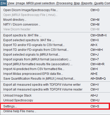
Current version 3.0.1 Alpha File menu screenshot, including the NIfTI / DICOM conversion and Settings items.
 spectrIm-QMRS
Version 3.0.1 Alpha
spectrIm-QMRS
Version 3.0.1 Alpha
Clinical Viewer GUI
The File menu contains the main commands for loading clinical DICOM MRS(I) data, converting scalar images between NIfTI and DICOM, importing and exporting spectra, connecting external result files to loaded datasets, and clearing the currently loaded image or spectroscopy data.
In routine use, the most important File menu action is usually Mount directory.... It lets spectrIm-QMRS scan a patient or study directory, identify image and spectroscopy series, and make these data available in the Patient/Study Browser. The new NIfTI / DICOM conversion... item opens the image conversion dialog for moving scalar image volumes between NIfTI files and DICOM image series.
For the clinical background behind DICOM import and DICOM formatted spectroscopy results, see the DICOM crash course.
Individual files can also be loaded directly. Export and import commands are intended for exchange with MATLAB, jMRUI, MIDAS, TDFDFit, CSV-based workflows, and external prediction or quantification tools.
Current version 3.0.1 Alpha File menu screenshot, including the NIfTI / DICOM conversion and Settings items.
Opens a single DICOM file from the local file system. This is useful for direct inspection or when only one image or spectroscopy object needs to be loaded.
Opens a spectroscopy file in jMRUI format. The older website noted that this function was disabled in the then-current version.
To be defined: confirm whether this command is active in version 3.0.1 Alpha and which jMRUI variants are supported.
Mounts a directory containing DICOM data. spectrIm-QMRS scans the selected directory and its subdirectories, detects image and spectroscopy series, and builds a patient/study/series tree for browsing clinical cases. This is the preferred clinical workflow: instead of selecting isolated files, the application can organize a complete examination and make the relevant DICOM objects directly accessible in the patient and study browser.
Opens a DICOM header viewer for the selected or loaded DICOM object. This is useful for checking acquisition parameters, private tags, spectroscopy-specific metadata, patient/study identifiers, and geometry information used to place MRS(I) data in anatomical context.
New item: add a current screenshot of the DICOM header viewer and document the available display/search/export options.
Opens the bidirectional image-conversion window. NIfTI-to-DICOM
conversion is handled inside spectrIm-QMRS for scalar
.nii and .nii.gz image volumes.
DICOM-to-NIfTI conversion scans DICOM image series and calls
the external dcm2niix executable.
Exports the loaded spectra to MATLAB .MAT format for further numerical analysis or visualization outside spectrIm-QMRS.
Exports only the currently selected spectra to MATLAB .MAT format. This is useful when analysis should be restricted to a region of interest or a selected voxel group.
Writes time-domain and/or frequency-domain signal data to CSV format for inspection, statistics, scripting, or exchange with external tools. CSV export is useful when spectra should be processed in R, Python, MATLAB, spreadsheet software, or an external machine-learning pipeline while preserving the link to the original voxel positions.
Exports selected spectra in jMRUI-compatible format. The voxel association string is used to preserve the link between spectra and their MRSI grid location. This makes it possible to process or inspect selected spectra in jMRUI while keeping enough information to associate later results with the original spectrIm-QMRS dataset.
Saves quantification results in jMRUI format so they can be reviewed or further processed in jMRUI-compatible workflows. For MRSI data, the voxel position association string is written with individual voxel results so that exported values can be traced back to their position in the original grid.
Exports measured spectra using the TDFDFit volume writer. This is intended for the newer volume-oriented TDFDFit workflow and parallel fitting pipeline.
To be defined: document the produced file set, naming scheme, and how this export connects to pthread-TDFDFit fitting.
Imports spectra from CSV files and associates rows with voxels
using the spectrIm-QMRS voxel position association string. In
the CSV file, each row represents one complex-valued spectrum
or signal result, and the first field should identify the voxel
position. The association string has the form x_y_z.
For example, 12_17_0 identifies the voxel at x=12,
y=17, z=0. For 2D MRSI data, z is normally 0. This convention
is used throughout spectrIm-QMRS to attach external data to a
specific spectrum in a multidimensional dataset.
Imports jMRUI-formatted spectra and associates them with the
already loaded dataset. The jMRUI data must contain position
identifiers that can be matched to spectrIm-QMRS voxel
coordinates. As with CSV import, the association string should
identify the voxel position in x_y_z form, counted from
the upper-left voxel of the MRSI grid. This allows externally
prepared spectra to be displayed and analyzed in their correct
anatomical and grid location.
Imports quantification results from jMRUI and associates the values with spectra or voxels in the currently loaded dataset. This command is intended for workflows where fitting or quantification has been performed outside spectrIm-QMRS, but the resulting metabolite values should be mapped back to the loaded MRS(I) dataset for display, comparison, or reporting.
Imports externally generated AI or machine-learning prediction results from CSV format. This can be used when another tool has classified spectra, predicted quality labels, estimated metabolite-related values, or produced other voxel-wise annotations. The first field should identify the voxel position so predictions can be mapped back to the loaded MRS(I) dataset.
To be defined: specify required column names, supported labels, and expected CSV format for version 3.0.1 Alpha.
Imports preprocessed MIDAS EPSI data. A geometrically matching DICOM spectroscopic image stack should be loaded first, so the imported EPSI data can be displayed in the correct examination coordinate system. spectrIm-QMRS uses the DICOM geometry, including slice position and orientation, to place the imported EPSI data correctly relative to anatomical image series from the same examination. After this association has been made, high-resolution MR images from the examination can be used as anatomical backgrounds for visual inspection of spectra.
Imports measured spectra using the TDFDFit volume reader. This complements the TDFDFit volume writer and supports workflows in which measured spectra are exchanged with the fitting engine.
To be defined: document the expected volume file format and typical use case.
Removes the currently loaded anatomical image stack from the viewer. Loaded spectroscopy data can remain available.
Removes the currently loaded spectroscopy data. The anatomical image stack can remain visible if it was loaded separately.
Opens the spectrIm-QMRS settings. These settings configure application behavior, reports, and processing pipelines. The default preprocessing action can be inspected and edited here. In version 3.0.1 Alpha the standard default preprocessing action performs HLSVDPro water removal, frequency alignment, and frequency-domain offset removal, with the offset range adapted to the loaded spectrum.