View menu
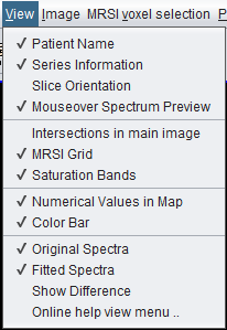
Version 3.0.1 Alpha View menu screenshot.
 spectrIm-QMRS
Version 3.0.3 Alpha International
spectrIm-QMRS
Version 3.0.3 Alpha International
Current release: 3.0.3 alpha international for Windows and Linux. This guide retains earlier screenshots and version-specific descriptions. Their original version labels are preserved; appearance and window placement may differ in 3.0.3. See the release notes.
Clinical Viewer GUI
The View menu controls which patient, series, image-overlay, map, and spectrum display elements are visible in the Clinical Viewer. Most entries are toggle items: selecting them turns a display layer or annotation on or off.
The View menu is mainly used to reduce visual clutter or to show additional context while reviewing MRS(I) data. During clinical review, users may want patient and series information visible. For screenshots, teaching, or anonymized presentations, these labels can be hidden.
Other toggles control overlays in the anatomical image panel and display options for spectral and metabolite map visualization. These settings do not change the underlying data; they only affect how loaded data are displayed.
Version 3.0.1 Alpha View menu screenshot.
Shows or hides the patient name in the viewer. This is useful during clinical review, but should normally be turned off when preparing anonymized screenshots, teaching material, or public documentation.
Shows or hides information about the currently loaded series, such as the series description or other display text derived from the DICOM data.
Shows or hides orientation information for the loaded MRS(I) dataset. This helps users understand how the spectroscopy grid is positioned relative to the anatomical image stack.
Enables a spectrum preview while the mouse pointer is moved over the MRSI grid. This gives a quick impression of spectral quality and signal content without explicitly selecting each voxel.
Shows or hides intersection overlays in the main image panel. These overlays help relate the currently displayed image slice to other image or spectroscopy geometry.
To be defined: add a more precise description once the current intersection overlay behavior is documented with screenshots.
Shows or hides the spectroscopy voxel grid on top of the anatomical image. The grid is central for selecting spectra and for understanding where each spectrum was measured.
Shows or hides saturation band overlays when this information is available in the loaded data. The previous documentation noted that this display works best for Siemens MRS(I) data.
Shows or hides numerical values inside the voxels of a displayed metabolite or parameter map. This can be helpful for inspecting exact values, but may be hidden when the map should be read mainly by color or when values overlap visually.
Shows or hides the color bar associated with a displayed map. The color bar helps interpret the scale used for metabolite, quality, or parameter overlays.
Shows or hides the measured spectra in the spectrum display panel. Keeping the original spectra visible is important when reviewing preprocessing and fitting results.
Shows or hides fitted spectra when fitting results are available. This allows users to compare measured data with the TDFDFit or other model fit.
Shows or hides the difference spectrum between the selected blue and red voxel groups. This is useful for comparing spectra from different anatomical regions, tissue classes, or selected voxel groups.