The ILV GUI
The current v3 alpha GUI is organized into four main tabs. They
separate the metabolite spin-system library, the pulse sequence
library, the simulation preview settings, and the basis-set batch
generation workflow.
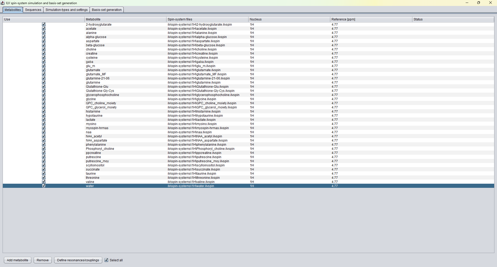
The Metabolites tab lists the available spin systems. Each row
connects a metabolite name with an ILV spin-system file, the
observed nucleus, a reference frequency in ppm, and a status field.
The user can include or exclude metabolites, add or remove entries,
and open the resonance/coupling editor. The spin-system definition
is where chemical shifts, relative spin amplitudes, and J-coupling
structure are defined before simulation.
Sequences tab
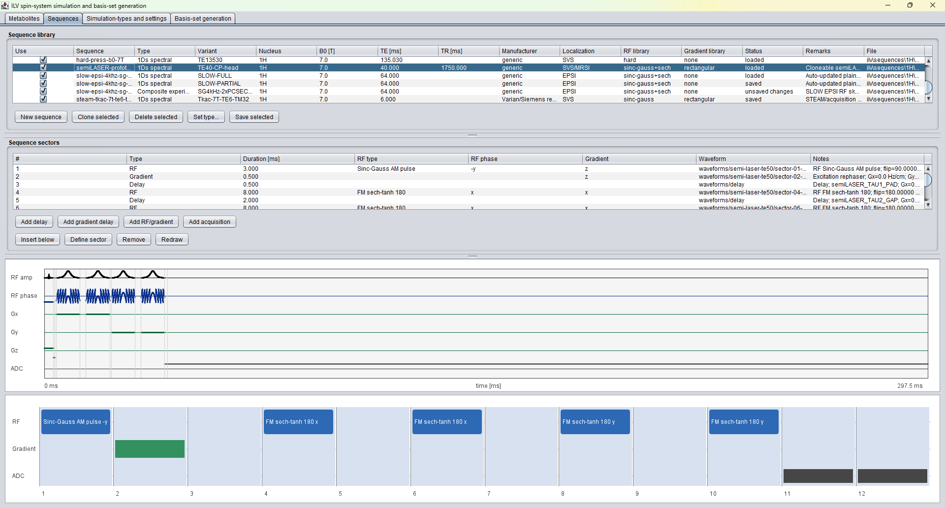
The Sequences tab is the graphical pulse sequence workspace. The
upper table describes complete sequence definitions, including
sequence type, variant, nucleus, B0, TE, TR, manufacturer,
localization type, RF library, gradient library, status, remarks,
and file path. The sector table below describes the individual
building blocks of the selected sequence: RF pulses, delays,
gradient delays, RF-gradient sectors, and acquisition sectors.
The lower part of the tab redraws the sequence timing as RF
amplitude, RF phase, gradient channels, and ADC activity. This is
the expert area for creating new sequences, cloning existing
definitions, inserting sectors, defining sector details, and saving
the sequence file.
Simulation-types and settings tab
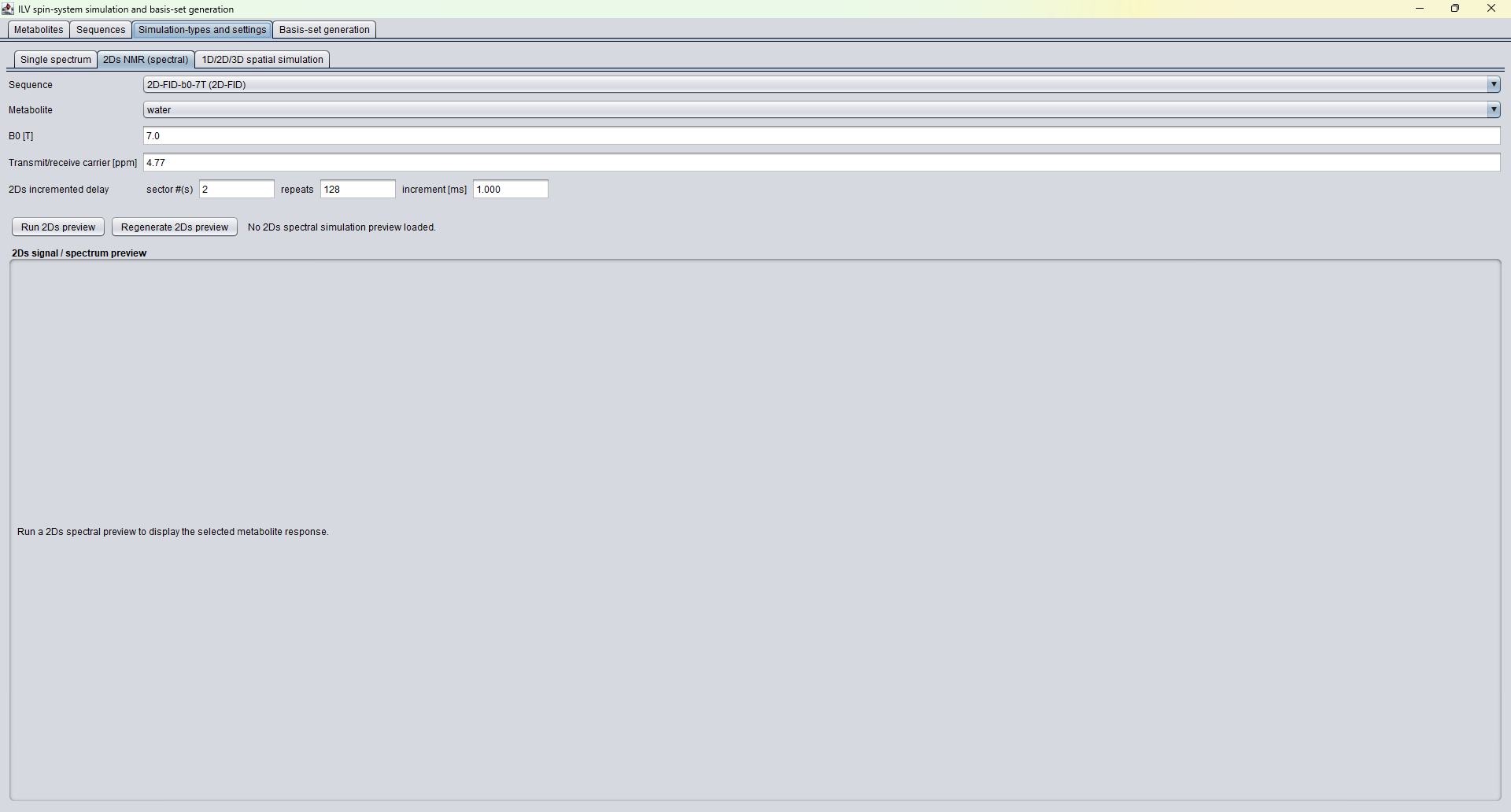
This tab selects how ILV should be run before a full basis-set
generation. It contains sub-tabs for single-spectrum simulation,
2D spectral simulation, and 1D/2D/3D spatial simulation. The
displayed example shows a 2D spectral simulation setup with the
selected sequence, metabolite, B0, transmit/receive carrier, and
the incremented delay settings. Preview buttons allow the user to
run or regenerate a simulation preview before committing to a full
basis-set calculation.
Single spectrum subtab
The Single spectrum subtab is used for one metabolite and one
sequence at a single spatial point. It is the fastest way to test
whether a spin-system definition, RF pulse, carrier frequency, and
acquisition setup produce the expected time-domain response and
spectrum. The controls select the sequence, metabolite, B0 field,
carrier position in ppm, transmit-carrier offset in Hz, and B1+
amplitude factor.
A sensitivity sweep can be run over either the B1+ amplitude factor
or the transmit offset. This is important for sequence development:
it lets the user inspect how robust a simulated metabolite response
is to RF-amplitude scaling or off-resonance excitation. In the ILV
parity work this kind of check was used as a practical guardrail:
before a basis set is trusted, a single spin system and sequence
should behave sensibly when the obvious experimental parameters are
perturbed.
The same subtab also contains the current SLOW editing preview
controls. SLOW editing uses two adiabatic 2pi pulse branches,
labelled FULL and PARTIAL in the GUI. The user can define the
center position, mhu, and beta for both branches, inspect the
calculated bandwidth in ppm, and show the RF-pulse effect as a
single spin-1/2 Bloch simulation from Mz=+1. The preview route
generates FULL, PARTIAL, and DIFFERENCE responses. This is a
research-level tool for developing and checking edited sequence
behaviour before those responses are used as fitting basis
functions.
2Ds NMR (spectral) subtab
The 2Ds NMR subtab is meant for explicit two-dimensional spectral
simulations. It uses a sequence that contains an incrementable
delay sector. The user selects the sequence and metabolite, sets
B0 and the transmit/receive carrier, and then defines which sector
or sectors are incremented, how many repeats are simulated, and the
delay increment in milliseconds.
Conceptually, this subtab asks: what happens to the simulated
signal when a specific evolution period in the sequence is stepped?
This is useful for J-resolved and other two-dimensional NMR style
experiments, and it is also useful when checking whether a sequence
definition is internally consistent. The preview makes it possible
to inspect the selected metabolite response before running a larger
basis-set generation.
In practical terms, this subtab is an expert diagnostic layer. It
is not the default clinical route, but it helps verify the sequence
sector model that the clinical route depends on. If the incremented
delay, carrier, or B0 is wrong, the resulting basis patterns will
not describe the measured signal correctly.
1D/2D/3D spatial simulation subtab
The spatial simulation subtab extends the ILV calculation from a
single spectrum to a grid of spatial positions. The user selects a
spatial sequence and metabolite, sets B0 and the carrier, and then
defines the X, Y, and Z simulation grid by start position, step
size, and number of points in centimeters.
This mode is designed to evaluate the spatial behaviour of a pulse
sequence: slice profiles, localization behaviour, transition
regions, and spatially dependent density matrices. The current GUI
can run the spatial density-matrix simulation, then calculate and
display expectation-value profiles or maps after the user selects
an operator. The displayed output can be shown as a 1D profile or
as a 2D image with horizontal and vertical cross-sections. The 3D
display is marked in the GUI as a future extension.
This is the natural place for expert validation of localization
sequences. For example, the semiLASER prototype work in the ILV
parity thread separated two questions that are easy to confuse:
first, whether the first ADC sample occurs at the declared TE;
second, whether the refocusing-pulse timing and gradient moments
are physically balanced. Spatial simulation gives a way to inspect
those sequence-design consequences before the same sequence is
used to generate a metabolite basis set for clinical data.
Basis-set generation tab
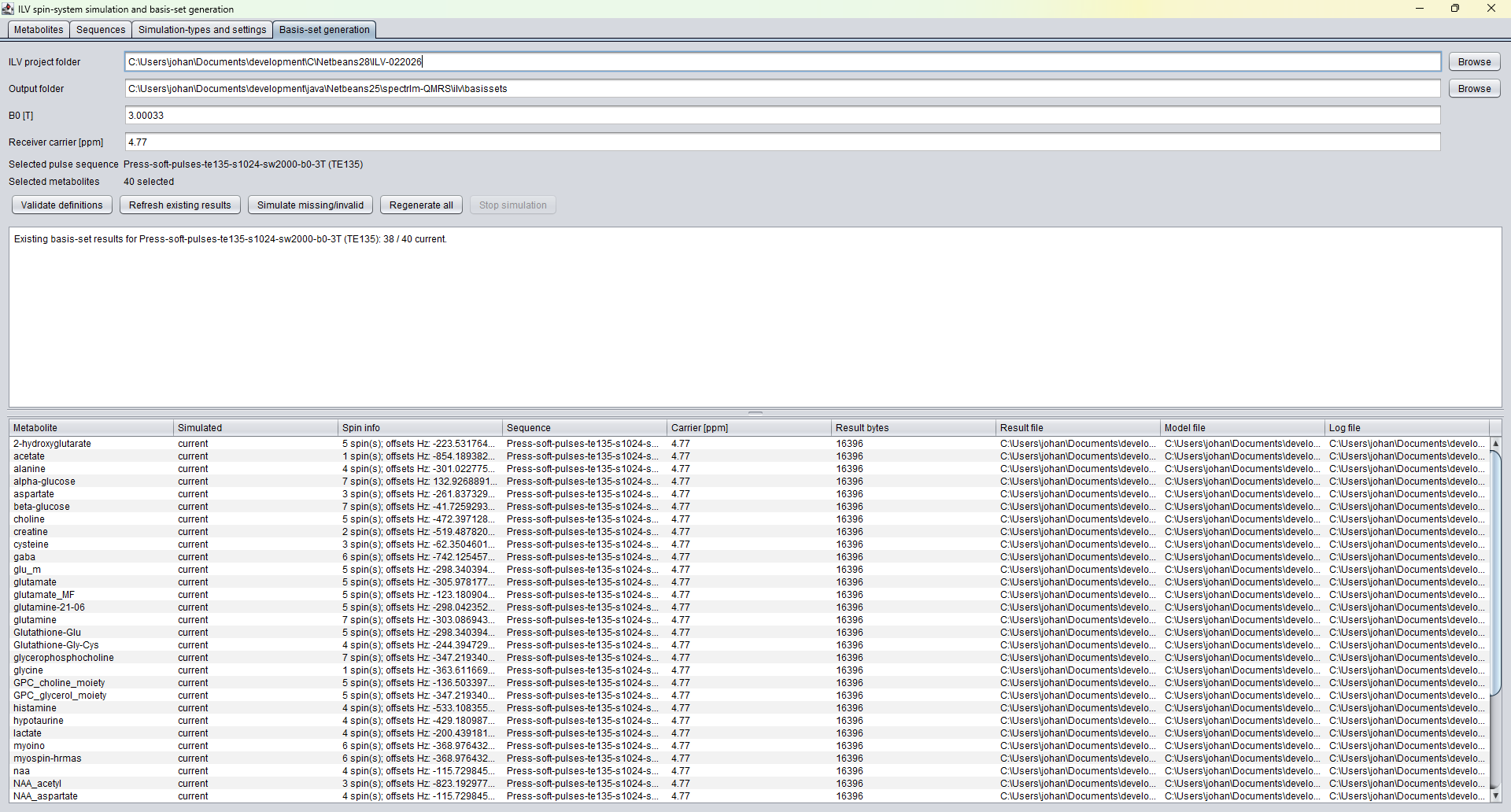
The Basis-set generation tab turns the selected definitions into a
set of simulated metabolite responses. It shows the ILV project
folder, the output folder, B0, receiver carrier, selected sequence,
and number of selected metabolites. The validation and refresh
buttons check whether existing simulations are current, missing, or
invalid. The simulation controls can generate missing/invalid basis
spectra or regenerate all spectra. The result table stores the
metabolite name, simulation status, spin information, sequence,
carrier, result byte count, result file, model file, and log file.
For SLOW-EPSI editing studies, the generated output can include both
the complete metabolite response and resonance-group components. The
resonance-group files use compact names such as
gaba_slow_full_3.003_ppm_td.bin, so the resulting
TDFDFit model can include a whole metabolite, selected multiplets, or
a mixture of both. This is useful when a coupled spin system contains
spectral regions that should be modelled with separate degrees of
freedom.