From the File menu
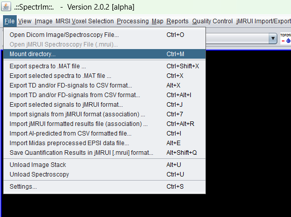
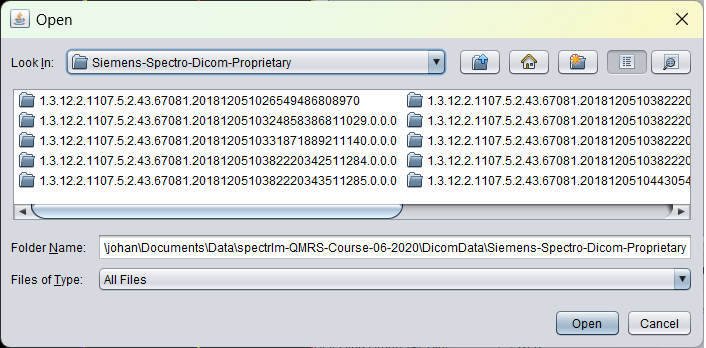
Use File > Mount directory... to select a directory containing a DICOM examination. spectrIm-QMRS scans the directory tree and builds the browser hierarchy from the discovered DICOM metadata.
 spectrIm-QMRS
Version 3.0.3 Alpha International
spectrIm-QMRS
Version 3.0.3 Alpha International
Current release: 3.0.3 alpha international for Windows and Linux. This guide retains earlier screenshots and version-specific descriptions. Their original version labels are preserved; appearance and window placement may differ in 3.0.3. See the release notes.
Clinical Viewer GUI
The Patient/Study Browser is the practical entry point for navigating mounted DICOM examinations in spectrIm-QMRS. It organizes patient, study, series, and instance information so that anatomical images and spectroscopy data can be selected without manually opening individual DICOM files.
The role of DICOM studies, series, instances, and UIDs in the clinical workflow is summarized in the DICOM crash course.
In the current workflow the browser is mainly used after mounting a DICOM directory tree from the File menu. It lets the user inspect the discovered DICOM hierarchy and select the image or spectroscopy series that should be loaded into the Clinical Viewer.
The same concept is important for the planned clinical Patient Browser described in the Clinical Cases menu. For clinical use, the browser should evolve from directory navigation into a patient-database view for reviewing already processed MRS(I) results, fitted spectra, metabolite maps, quality indicators, and reports.
The browser occupies the lower part of the Clinical Viewer. The left tree shows mounted DICOM directories and discovered patient/study structure. The right table lists DICOM instances from the selected series, including metadata such as file name, type, instance number, slice position, field of view, echo time, and repetition time.
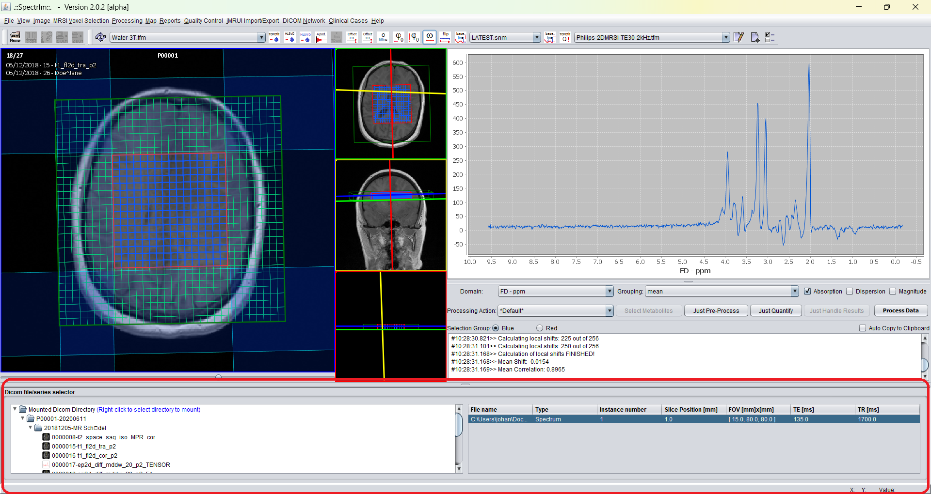
This screenshot is from the previous website and is kept because it documents the browser concept well. Replace it with a current version 3.0.1 Alpha screenshot once a clean documentation dataset is available.
Use File > Mount directory... to select a directory containing a DICOM examination. spectrIm-QMRS scans the directory tree and builds the browser hierarchy from the discovered DICOM metadata.
The toolbar mount button provides the same entry point for routine work. After a directory is mounted, the patient/study tree can be expanded and the relevant image or spectroscopy series can be selected.
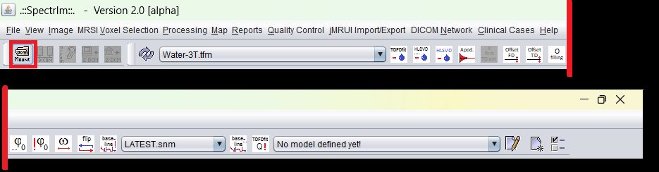
Once mounted, the selected DICOM directory appears in the browser tree. Expanding the tree reveals the available patient/study/series entries. Selecting a series populates the instance table on the right; selecting or opening an instance loads the corresponding image or spectroscopy data into the viewer.
The future clinical Patient Browser should provide a higher-level view than a raw DICOM directory browser. Instead of only showing files and series, it should help clinicians navigate patient cases, studies, processed MRS(I) datasets, fitting models, quantitative maps, quality-control labels, and generated reports.
This is especially important when spectrIm-QMRS is used in hospital environments where installation of Java or Python components is not permitted. Version 3.0.1 Alpha already supports a self-contained ZIP package; a clinical browser would complement that by making database-oriented review more direct for MD users.
For testing and training, the website provides anonymized Siemens and Philips spectroscopy DICOM example datasets. From version 3.1 alpha onward, United Imaging spectroscopy file-format support is planned as an additional input route.
Loading, mounting, importing, exporting, and unloading DICOM image or spectroscopy data.
Download anonymized Siemens and Philips spectroscopy DICOM examples for testing the browser and loading workflow.
Planned patient browser direction for clinical MRS(I) result navigation.
Written workflow for loading data, inspecting spectra, preprocessing, and creating simple spectroscopic maps.