Clinical Introduction to MRS(I)
A clinician-friendly overview of what MRS and MRSI measure, why quality control matters, and how processing connects spectra to metabolite maps. Start with the introduction.
 spectrIm-QMRS
Version 3.0.3 Alpha International
Version 3.0.3 Alpha International
spectrIm-QMRS is a desktop application for viewing, processing, modeling and quantifying magnetic resonance spectroscopy and spectroscopic imaging data.
spectrIm-QMRS 3.0.3 alpha international is now available for Windows 11 x64 and Linux x64. Both packages include their own Java and Python runtimes, NumPy and SciPy, and platform-specific ILV simulation and pthreads TDFDFit executables. Extract the complete package and use its startup script.
Still an alpha release. This version has been thoroughly tested in our workflows on Windows 11 and Ubuntu 22.04 under WSLg, but bugs may remain. Other Linux environments have not been verified to the same extent. Back up your data and models, check processing results carefully, and report problems with the relevant logs.
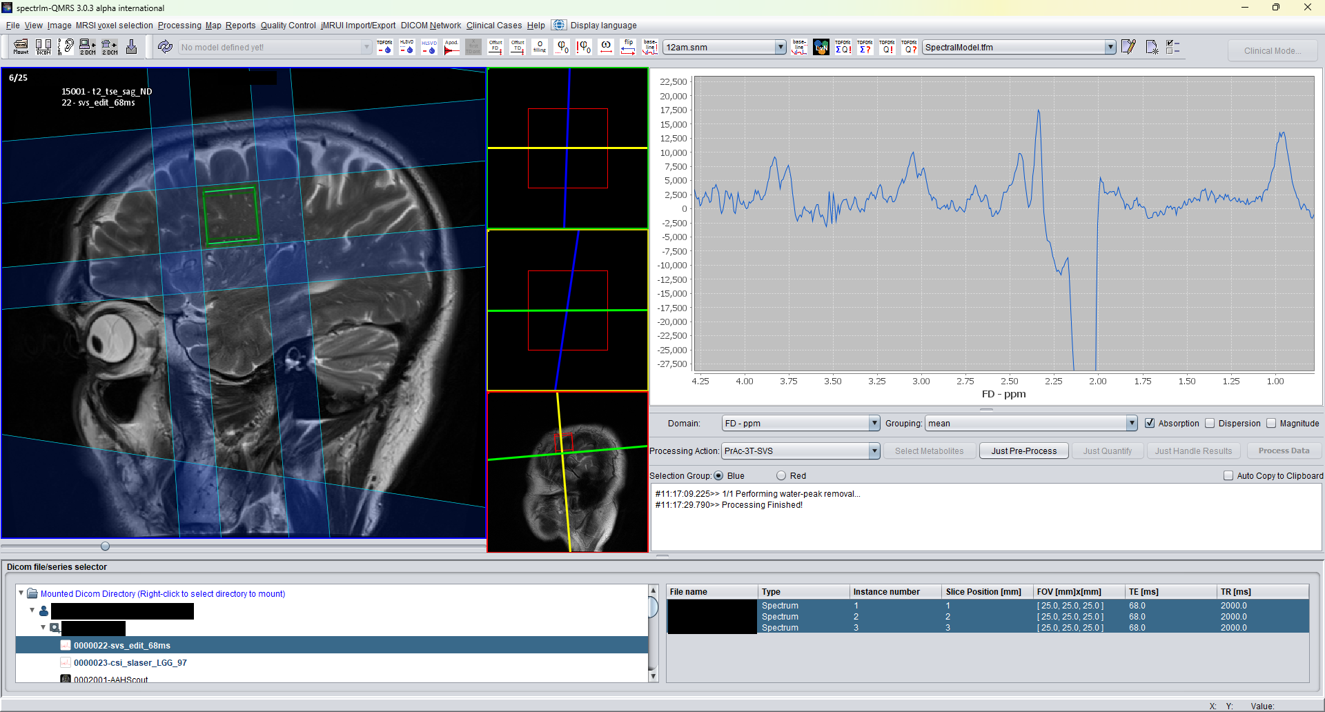
New users can start with the main menu and toolbar overview. It explains how the Clinical Viewer is organized and links to the detailed v3 documentation pages for each menu.
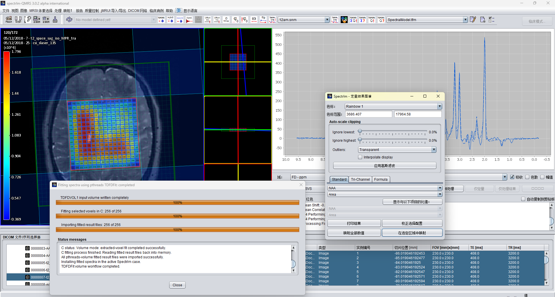
The international interface lets users select the display language while keeping the same clinical MRS(I) workflow and tools. The example below shows the Clinical Viewer in Simplified Chinese.
A clinician-friendly overview of what MRS and MRSI measure, why quality control matters, and how processing connects spectra to metabolite maps. Start with the introduction.
Distributed as self-contained Windows ZIP and Linux TAR.GZ packages including the required Java and Python runtime components.
The interface is available in English, Arabic, Chinese (Simplified), Czech, Danish, Dutch, Finnish, French, German, Hindi, Icelandic, Italian, Japanese, Norwegian Bokmål, Polish, Portuguese, Spanish, and Swedish. See the Chinese interface.
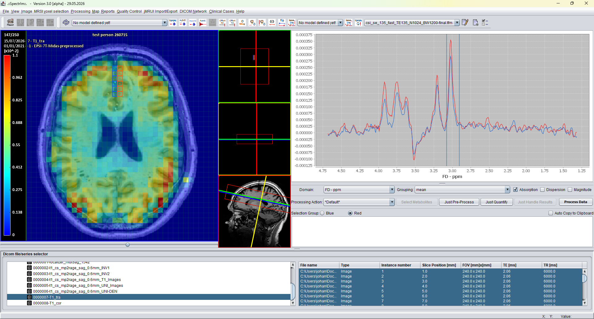
A more advanced HLSVD implementation provides fast residual water removal, denoising, and preprocessing of clinical MRS(I) datasets. See the interactive processing workflow.
Three-dimensional CSI and MRSI datasets can now be loaded from multiframe DICOM spectroscopy objects and displayed with their full spatial geometry.
Enhanced spectroscopy DICOM from Siemens XA software lines is accepted as valid input, while the legacy Siemens spectroscopy DICOM format remains supported.
Integrated ILV simulation solves the Liouville-von Neumann equations to generate quantum-mechanical metabolite basis spectra and multiplet-resolved components for prior-knowledge model construction. Open the simulator page.
SLOW FULL, PARTIAL, and DIFFERENCE datasets can be analysed with ILV-generated basis spectra, including shorter component names and resonance-group components for more flexible TDFDFit modelling. Open the SLOW-EPSI page.
Guided prior knowledge model creation to help clinicians start working with MRS(I) without requiring a spectroscopist as a direct colleague.
A two-water-compartment model supports LCModel-style absolute quantification of SVS data using a same-study water reference and compartment-corrected concentration estimates. Open the absolute quantification page.
TDFDFit now performs prior-knowledge fitting in parallel with POSIX threads for faster processing across available CPU cores. Apply predefined models.
Clinicians can select a region of voxels, fit the summed
spectrum as one higher-SNR spectrum with ΣQ!,
and reopen named summed regions with Σ? to
review the selected voxels, fitted overlay, and fitted
parameters.
Read the processing menu notes.
A manual conversion window now supports scalar NIfTI-to-DICOM image-series creation and DICOM-to-NIfTI conversion through dcm2niix. Open the conversion page.
A new crash course explains why DICOM receiving and DICOM formatted spectroscopy results matter in clinical MRS(I), and how this makes spectrIm-QMRS more than a standalone research processing tool. Read the crash course.
Clinical Viewer screenshots and selected local image files can be written as DICOM Secondary Capture objects using the last loaded spectroscopy DICOM examination as patient and study context. Open the workflow page.
From version 3.1 alpha onward, spectrIm-QMRS is planned to add support for United Imaging spectroscopy file formats alongside the existing DICOM-based Siemens and Philips example workflows.
Prior-knowledge model construction for NMR/MR spectroscopy knowledgeable users: Voigt-line editing, prototype optimization, and parameter-linking documentation. Open the modelling frame page.
Legacy expert route for loading externally simulated MRUI numeric basis patterns into TDFDFitControl. In v3 this route is complemented by the integrated ILV basis-set simulator. Open the walkthrough.